3.3 Organelles of Eukaryotic Cells
Learning Objectives
By the end of this section, you will be able to:
- Describe the structure of eukaryotic plant and animal cells
- Summarize the differences among the components of prokaryotic cells, animal cells, and plant cells
- Summarize the functions of the plasma membrane and major cell organelles
Watch a video about cellular organelles.
Have you ever heard the phrase “form follows function”? It’s a philosophy that many industries follow. In architecture, this means that buildings should be constructed to support the activities that will be carried out inside them. For example, a skyscraper should include several elevator banks. A hospital should have its emergency room easily accessible.
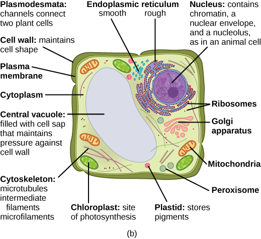
Our natural world also utilizes the principle of form following function, especially in cell biology, and this will become clear as we explore eukaryotic cells (Figure 3.7). Unlike prokaryotic cells, eukaryotic cells have: 1) a membrane-bound nucleus; 2) numerous membrane-bound organelles such as the endoplasmic reticulum, Golgi apparatus, chloroplasts, mitochondria, and others; and 3) several, rod-shaped chromosomes. Because a membrane surrounds eukaryotic cell’s nucleus, it has a “true nucleus.” The word “organelle” means “little organ,” and, as we already mentioned, organelles have specialized cellular functions, just as your body’s organs have specialized functions.
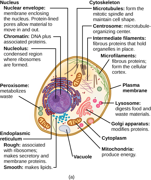
Like prokaryotes, eukaryotic cells have a plasma membrane, the cell membrane, made up of a phospholipid bilayer with embedded proteins that separates the internal contents of the cell from its surrounding environment. A phospholipid is a lipid molecule composed of two fatty acid chains, a glycerol backbone, and a phosphate group. The plasma membrane is selectively permeable. This means that the membrane allows some materials to freely enter or leave the cell, while other materials cannot move freely, but require a specialized structure, and occasionally, even energy investment for crossing. Plasma membranes are asymmetric. On the membrane’s interior, some proteins serve to anchor the membrane to the cytoskeleton’s fibres. There are peripheral proteins on the membrane’s exterior that bind extracellular matrix elements.
The cytoskeleton has three different types of protein elements. Microfilaments provide rigidity and shape to the cell, and facilitate cellular movements. Intermediate filaments bear tension and anchor the nucleus and other organelles in place. Microtubules help the cell resist compression, serve as tracks for motor proteins that move vesicles through the cell, and pull replicated chromosomes to opposite ends of a dividing cell. Animal cells may have cilia and flagella that mediate cell movement (not shown in Figure 3.7a).
Unlike the animal cell (Figure 3.7a), the plant cell (Figure 3.7b) has a structure external to the plasma membrane called the cell wall. The cell wall is a rigid covering that protects the cell, provides structural support, and gives shape to the cell. Fungal and protist cells also have cell walls. A plasmodesma is a channel between the cell walls of two adjacent plant cells. Animal cells can be physically joined by tight junctions, gap junctions, and desmosomes. Plant cells also have specialized storage compartments, plastids and the large central vacuole, that are absent in animal cells.
Eukaryotic cells obtain most of their energy-carrying molecule in the form of ATP from cellular respiration occurring in mitochondria, often called the “powerhouses” or “energy factories” of a cell. In keeping with our theme of form following function, it is important to point out that muscle cells have a very high concentration of mitochondria because muscle cells need a lot of energy to contract. Plant cells have mitochondria but also chloroplasts. Photosynthesis takes place in chloroplasts.
The endomembrane system consists of the nuclear envelope, endoplasmic reticulum, Golgi apparatus, vesicles, vacuoles, and lysosomes what work together to produce, modify, package, and transport lipids and proteins. The plasma membrane interacts with the endomembranous organelles and is part of the endomembrane system. The rough endoplasmic reticulum is associated with ribosomes and makes proteins. Smooth endoplasmic reticulum is not attached to ribosomes and synthesize lipids. The Golgi apparatus chemically modifies lipids and proteins and packages the material for distribution. In fact, the Golgi has two distinct sides to the structure, namely the cis-face and the trans-face. The cis-face is named because this side of the Golgi faces the centre of the cell and receives the biosynthetic output from the rough endoplasmic reticulum. The output from the Golgi is contained in vesicles at the trans-face that faces the cell membrane. Vesicles and vacuoles are membrane-bound sacs that function in storage and transport. Lysosomes contain digestive enzymes. The endomembrane system does not include either mitochondria or chloroplast membranes or peroxisomes.
The details of cellular components are covered in subsequent sections:
- Endomembrane system, vesicles, vacuoles, endoplasmic reticulum, Golgi apparatus, and lysosomes (Chapter 3.4)
- Cytoskeleton (Chapter 3.5)
- Connections between cells (Chapter 3.6)
- Mitochondria and chloroplasts (Chapter 3.7)
At this point, it should be clear that eukaryotic cells have a more complex structure than do prokaryotic cells. Organelles allow for various functions to occur in the cell at the same time. Before discussing the functions of organelles within a eukaryotic cell, let us first examine two important compartments of the cell: the cytoplasm and the nucleus.
The Cytoplasm
The cytoplasm comprises the contents of a cell between the plasma membrane, also called the cell membrane, and the nuclear envelope (a structure to be discussed shortly). It is made up of organelles suspended in the gel-like cytosol, the cytoskeleton, and various chemicals. Even though the cytoplasm consists of 70 to 80 percent water, it has a semi-solid consistency, which comes from the proteins within it. However, proteins are not the only organic molecules found in the cytoplasm. Glucose and other simple sugars, polysaccharides, amino acids, nucleic acids, fatty acids, and derivatives of glycerol are found there too. Ions of sodium, potassium, calcium, and many other elements are also dissolved in the cytoplasm. Many metabolic reactions, including protein synthesis, take place in the cytoplasm.
The Nucleus
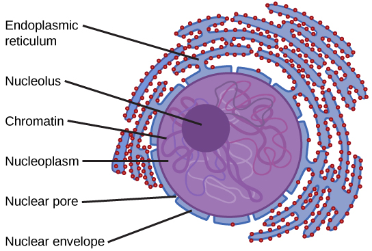
Typically, the nucleus is the most prominent organelle in a cell, not to be confused with the nucleus of an atom or ion. The nucleus (plural: nuclei) houses the cell’s DNA in the form of chromatin and directs the synthesis of ribosomes and nucleic acids for making proteins in the cytoplasm. The structure of the nucleus is shown in Figure 3.8. The nuclear envelope is a double-membrane structure that constitutes the outermost portion of the nucleus. Both the inner and outer membranes of the nuclear envelope are phospholipid bilayers. The nuclear envelope is punctuated with pores that control the passage of ions, molecules, and RNA between the nucleoplasm and the cytoplasm.
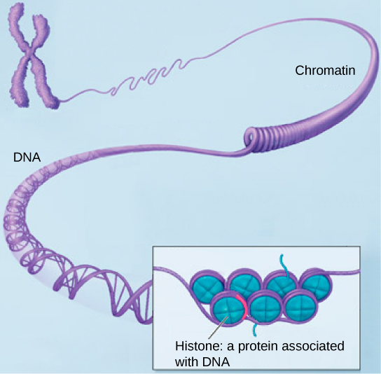
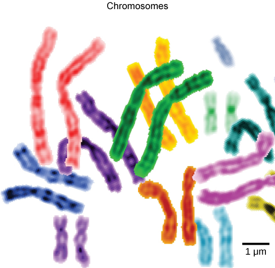
To understand chromatin, it is helpful to first consider chromosomes. Chromosomes are structures within the nucleus that are made up of DNA, the hereditary material, and histone proteins (Figure 3.9). This combination of DNA and histones is called chromatin. Chromatin is organized into chromosome. In eukaryotes, chromosomes are linear structures. Chromosomes are only visible and distinguishable from one another when the cell is getting ready to divide. When the cell is in the growth and maintenance phases of its life cycle, the chromosomes resemble an unwound, jumbled bunch of threads.
Every species has a specific number of chromosomes in the nucleus of its body cells. For example, in fruit flies, the chromosome number is eight, whereas in humans, the chromosome number is 46. The 46 chromosomes can be grouped as 23 maternal-paternal pairs (Figure 3.10), where one set of 23 chromosomes is inherited from the mother and another set of 23 chromosomes is inherited from the father.
We already know that the nucleus directs the synthesis of ribosomes, but how does it do this? Some chromosomes have sections of DNA that encode ribosomal RNA. A darkly stained area within the nucleus, called the nucleolus (plural: nucleoli), aggregates the ribosomal RNA with associated proteins to assemble the ribosomal subunits that are then transported through the nuclear pores into the cytoplasm.
CAREER CONNECTION
Geneticist
Many diseases arise from genetic mutations that prevent synthesizing critical proteins. One such disease is Lowe disease (or oculocerebrorenal syndrome, because it affects the eyes, brain, and kidneys). In Lowe disease, there is a deficiency in an enzyme localized to the Golgi apparatus. Children with Lowe disease are born with cataracts, typically develop kidney disease after the first year of life, and may have impaired mental abilities.
A mutation on the X chromosome causes Lowe disease. The X chromosome is one of the two human sex chromosomes, as these chromosomes determine a person’s sex. Females possess two X chromosomes while males possess one X and one Y chromosome. In females, the genes on only one of the two X chromosomes are expressed. Females who carry the Lowe disease gene on one of their X chromosomes are carriers and do not show symptoms of the disease. However, males only have one X chromosome and the genes on this chromosome are always expressed. Therefore, males will always have Lowe disease if their X chromosome carries the Lowe disease gene. Geneticists have identified the mutated gene’s location, as well as many other mutation locations that cause genetic diseases. Through prenatal testing, a woman can find out if the fetus she is carrying may be afflicted with one of several genetic diseases.
Geneticists analyze prenatal genetic test results and may counsel pregnant women on available options. They may also conduct genetic research that leads to new drugs or foods, or perform DNA analyses for forensic investigations.
Ribosomes
Ribosomes are the cellular structures responsible for protein synthesis. When viewed through an electron microscope, free ribosomes appear as either clusters or single tiny dots floating freely in the cytoplasm. Ribosomes may be attached to either the cytoplasmic side of the plasma membrane or the cytoplasmic side of the endoplasmic reticulum. Electron microscopy has shown that ribosomes consist of large and small subunits. Ribosomes are complexes of protein and catalytic ribosomal RNA that are responsible for protein synthesis.
Because protein synthesis is essential for all cells, ribosomes are found in practically every cell, although they are smaller in prokaryotic cells. They are particularly abundant in immature red blood cells for the synthesis of hemoglobin, which functions in the transport of oxygen throughout the body.
Peroxisomes
Peroxisomes are small, round organelles enclosed by single membranes. They carry out oxidation reactions that break down fatty acids and amino acids. They also detoxify many poisons that may enter the body. Alcohol is detoxified by peroxisomes in liver cells. A byproduct of these oxidation reactions is hydrogen peroxide, H2O2, which is contained within the peroxisomes to prevent the chemical from causing damage to cellular components outside of the organelle. Hydrogen peroxide is safely broken down by peroxisomal enzymes into water and oxygen.
Animal Cells versus Plant Cells
Despite their fundamental similarities, there are some striking differences between animal and plant cells. Animal cells have centrosomes, centrioles, and lysosomes, whereas plant cells do not. Plant cells have cell wall, chloroplasts, plasmodesmata, plastids, and a large central vacuole, whereas animal cells do not.
The Centrosome
Found near the nuclei of animal cells, the centrosome is a microtubule-organizing centre from where all microtubules originate. The centrosome is a pair of centrioles, two structures that lie perpendicular to each other (Figure 3.11). Each centriole is a cylinder of nine triplets of microtubules.

The centrosome replicates itself before a cell divides, and the centrioles play a role in pulling the duplicated chromosomes to opposite ends of the dividing cell. However, the exact function of the centrioles in cell division is not clear, since cells that have the centrioles removed can still divide, and plant cells, which lack centrioles, are capable of cell division.
The Cell Wall
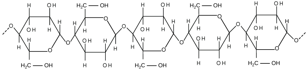
The cell wall, a rigid covering that protects the cell, provides structural support, and gives shape to the cell. Fungal and some protistan cells also have cell walls. While the prokaryotic cell walls’ chief component is peptidoglycan, the major organic molecule in the plant (and some protists’) cell wall is cellulose (Figure 3.12), a polysaccharide comprised of long, straight chains of glucose units. Have you ever noticed that when you bite into a raw vegetable, like celery, it crunches? That’s because you are tearing the celery cells’ rigid cell walls with your teeth. When nutritional information refers to dietary fibre, it is referring to the cellulose content of food.
The Central Vacuole
The central vacuole plays a key role in regulating the cell’s concentration of water in changing environmental conditions. In plant cells, the liquid inside the central vacuole provides turgor pressure, which is the outward pressure caused by the fluid inside the cell. Have you ever noticed that if you forget to water a plant for a few days, it wilts? That is because as the water concentration in the soil becomes lower than the water concentration in the plant, water moves out of the central vacuoles and cytoplasm and into the soil. As the central vacuole shrinks, it leaves the cell wall unsupported. This loss of support to the cell walls of a plant results in the wilted appearance. Additionally, this fluid has a very bitter taste, which discourages consumption by insects and animals. The central vacuole also functions to store proteins in developing seed cells. Furthermore, in plant cells, the vacuole expands, enlarging the cell without the need to produce more cytoplasm and also breaks down macromolecules.
Intercellular connnections
Animal cells communicate through their extracellular matrices and are physically connected to each other by tight junctions, gap junctions, and desmosomes. Plant cells are connected and communicate with each other by plasmodesmata.
LINK TO LEARNING
To conduct a virtual microscopy lab and review the parts of a cell, work through the steps of this interactive assignment.
Section Summary
The functions of cellular components are summarized in Table 3.1.
Table 3.1 Components of Prokaryotic and Eukaryotic Cells and Their Functions
| Cell Component | Function | Present in Prokaryotes? | Present in Animal Cells? | Present in Plant Cells? |
|---|---|---|---|---|
| Plasma membrane | Separates cell from external environment; controls passage of organic molecules, ions, water, oxygen, and wastes into and out of the cell | Yes | Yes | Yes |
| Cytoplasm | Provides structure to cell; site of many metabolic reactions; medium in which organelles are found | Yes | Yes | Yes |
| Nucleoid | Location of DNA | Yes | No | No |
| Nucleus | Cell organelle that houses DNA and directs synthesis of ribosomes and proteins | No | Yes | Yes |
| Ribosomes | Protein synthesis | Yes | Yes | Yes |
| Mitochondria | ATP production/cellular respiration | No | Yes | Yes |
| Peroxisomes | Oxidizes and breaks down fatty acids and amino acids, and detoxifies poisons | No | Yes | Yes |
| Vesicles and vacuoles | Storage and transport; digestive function in plant cells | No | Yes | Yes |
| Centrosome | Unspecified role in cell division in animal cells; organizing center of microtubules in animal cells | No | Yes | No |
| Lysosomes | Digestion of macromolecules; recycling of worn-out organelles | No | Yes | No |
| Cell wall | Protection, structural support and maintenance of cell shape | Yes, primarily peptidoglycan in bacteria but not Archaea | No | Yes, primarily cellulose |
| Chloroplasts | Photosynthesis | No | No | Yes |
| Endoplasmic reticulum | Modifies proteins and synthesizes lipids | No | Yes | Yes |
| Golgi apparatus | Modifies, sorts, tags, packages, and distributes lipids and proteins | No | Yes | Yes |
| Cytoskeleton | Maintains cell’s shape, secures organelles in specific positions, allows cytoplasm and vesicles to move within the cell, and enables unicellular organisms to move independently | Yes | Yes | Yes |
| Flagella | Cellular locomotion | Some | Some | No, except for some plant sperm. |
| Cilia | Cellular locomotion, movement of particles along extracellular surface of plasma membrane, and filtration | No | Some | No |
Exercises
Glossary
cell wall: a rigid cell covering made of cellulose in plants, peptidoglycan in bacteria, non-peptidoglycan compounds in Archaea, and chitin in fungi that protects the cell, provides structural support, and gives shape to the cell
central vacuole: a large plant cell organelle that acts as a storage compartment, water reservoir, and site of macromolecule degradation
chloroplast: a plant cell organelle that carries out photosynthesis
cilium (plural: cilia): a short, hair-like structure that extends from the plasma membrane in large numbers and is used to move an entire cell or move substances along the outer surface of the cell
cytoplasm: the entire region between the plasma membrane and the nuclear envelope, consisting of organelles suspended in the gel-like cytosol, the cytoskeleton, and various chemicals
cytoskeleton: the network of protein fibers that collectively maintains the shape of the cell, secures some organelles in specific positions, allows cytoplasm and vesicles to move within the cell, and enables unicellular organisms to move
cytosol: the gel-like material of the cytoplasm in which cell structures are suspended
desmosome: a linkage between adjacent epithelial cells that forms when cadherins in the plasma membrane attach to intermediate filaments
endomembrane system: the group of organelles and membranes in eukaryotic cells that work together to modify, package, and transport lipids and proteins
endoplasmic reticulum (ER): a series of interconnected membranous structures within eukaryotic cells that collectively modify proteins and synthesize lipids
extracellular matrix: the material, primarily collagen, glycoproteins, and proteoglycans, secreted from animal cells that holds cells together as a tissue, allows cells to communicate with each other, and provides mechanical protection and anchoring for cells in the tissue
flagellum (plural: flagella): the long, hair-like structure that extends from the plasma membrane and is used to move the cell
gap junction: a channel between two adjacent animal cells that allows ions, nutrients, and other low-molecular weight substances to pass between the cells, enabling the cells to communicate
Golgi apparatus: a eukaryotic organelle made up of a series of stacked membranes that sorts, tags, and packages lipids and proteins for distribution
lysosome: an organelle in an animal cell that functions as the cell’s digestive component; it breaks down proteins, polysaccharides, lipids, nucleic acids, and even worn-out organelles
mitochondria (singular: mitochondrion): the cellular organelles responsible for carrying out cellular respiration, resulting in the production of ATP, the cell’s main energy-carrying molecule
nuclear envelope: the double-membrane structure that constitutes the outermost portion of the nucleus
nucleolus: the darkly staining body within the nucleus that is responsible for assembling ribosomal subunits
nucleus: the cell organelle that houses the cell’s DNA and directs the synthesis of ribosomes and proteins
peroxisome: a small, round organelle that contains hydrogen peroxide, oxidizes fatty acids and amino acids, and detoxifies many poisons
plasma membrane: a phospholipid bilayer with embedded (integral) or attached (peripheral) proteins that separates the internal contents of the cell from its surrounding environment
plasmodesma (plural: plasmodesmata): a channel that passes between the cell walls of adjacent plant cells, connects their cytoplasm, and allows materials to be transported from cell to cell
ribosome: a cellular structure that carries out protein synthesis
rough endoplasmic reticulum (RER): the region of the endoplasmic reticulum that is studded with ribosomes and engages in protein modification
smooth endoplasmic reticulum (SER): the region of the endoplasmic reticulum that has few or no ribosomes on its cytoplasmic surface and synthesizes carbohydrates, lipids, and steroid hormones; detoxifies chemicals like pesticides, preservatives, medications, and environmental pollutants, and stores calcium ions
tight junction: a firm seal between two adjacent animal cells created by protein adherence
vacuole: a membrane-bound sac, somewhat larger than a vesicle, that functions in cellular storage and transport
vesicle: a small, membrane-bound sac that functions in cellular storage and transport; its membrane is capable of fusing with the plasma membrane and the membranes of the endoplasmic reticulum and Golgi apparatus
Media Attributions
- Figure 3.8 modification of work by NIGMS, NIH
- Figure 3.10 modification of work by NIH; scale-bar data from Matt Russell
