3.4 The Endomembrane System
Learning Objectives
By the end of this section, you will be able to:
- List the components of the endomembrane system
- Recognize the relationship between the endomembrane system and its functions
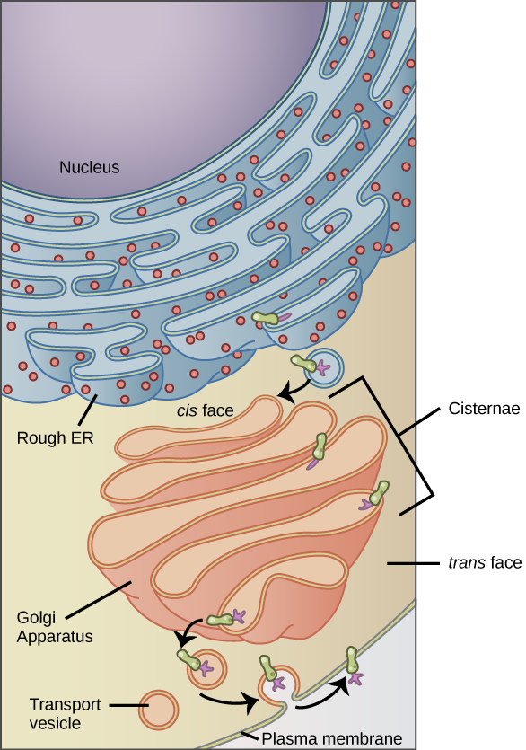
The endomembrane system (endo = “within”) is a group of membranes and organelles (Figure 3.13) in eukaryotic cells that works together to modify, package, and transport lipids and proteins. It includes the nuclear envelope, vesicles, vacuoles, endoplasmic reticulum, Golgi apparatus, and lysosomes. Although not technically within the cell, the plasma membrane is included in the endomembrane system because it interacts with the other endomembranous organelles. The endomembrane system does not include either mitochondria or chloroplast membranes or peroxisomes.
VISUAL CONNECTION
Vesicles and Vacuoles
Vesicles and vacuoles are membrane-bound sacs that function in storage and transport. Vacuoles are somewhat larger than vesicles, and the membrane of a vacuole does not fuse with the membranes of other cellular components. Vesicles can fuse with other membranes within the cell. Additionally, enzymes within plant vacuoles can break down macromolecules.
Endoplasmic Reticulum
The endoplasmic reticulum (ER) is a series of interconnected membranous tubules that collectively modify proteins and synthesize lipids. However, these two functions are performed in separate areas of the endoplasmic reticulum: the rough endoplasmic reticulum and the smooth endoplasmic reticulum, respectively. The hollow portion of the ER tubules is called the lumen or cisternal space. The membrane of the ER, which is a phospholipid bilayer embedded with proteins, is continuous with the nuclear envelope.
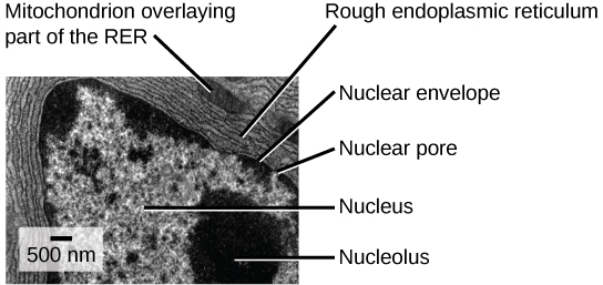
The rough endoplasmic reticulum (RER) is so named because the ribosomes attached to its cytoplasmic surface give it a studded appearance when viewed through an electron microscope (Figure 3.14). The ribosomes synthesize proteins while attached to the ER, resulting in the transfer of their newly synthesized proteins into the lumen of the RER where they undergo modifications such as folding or addition of sugars. The RER also makes phospholipids for cell membranes. If the phospholipids or modified proteins are not destined to stay in the RER, they will be packaged within vesicles and transported from the RER by budding from the membrane. Since the RER is engaged in modifying proteins that will be secreted from the cell, it is abundant in cells that secrete proteins, such as liver cells.
The smooth endoplasmic reticulum (SER) is continuous with the RER but has few or no ribosomes on its cytoplasmic surface. The SER’s functions include synthesis of carbohydrates, lipids (including phospholipids), and steroid hormones; detoxification of medications and poisons; alcohol metabolism; and storage of calcium ions. In muscle cells, a specialized SER, the sarcoplasmic reticulum, is responsible for storing calcium ions that are needed to trigger the muscle cells’ coordinated contractions.
Golgi Apparatus
We have already mentioned that vesicles can bud from the ER, but where do the vesicles go? Before reaching their final destination, the lipids or proteins within the transport vesicles need to be sorted, packaged, and tagged so that they wind up in the right place. The sorting, tagging, packaging, and distribution of lipids and proteins take place in the Golgi apparatus (also called the Golgi body), a series of flattened membranous sacs (Figure 3.15). In plant cells, the Golgi has an additional role of synthesizing polysaccharides, some of which are incorporated into the cell wall and some of which are used in other parts of the cell.
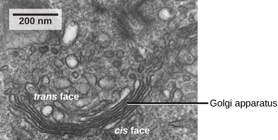
The Golgi apparatus has a receiving face, cis-face, near the ER and a releasing face, trans-face, on the side away from the ER, toward the cell membrane. The transport vesicles that form from the ER travel to the receiving face, fuse with it, and empty their contents into the lumen of the Golgi apparatus. As the proteins and lipids travel through the Golgi, they undergo further modifications, such as glycosylation. The most frequent modification is the addition of short chains of sugar molecules. The newly modified proteins and lipids are then tagged with phosphate groups or other small molecules to enable them to be routed to their proper destinations. Finally, the modified and tagged proteins are packaged into vesicles that bud from the opposite face of the Golgi. While transport vesicles deposit their contents into other parts of the cell where they will be used, secretory vesicles fuse with the plasma membrane and release their contents outside the cell.
The amount of Golgi in different cell types again illustrates that form follows function within cells. Cells that engage in a great deal of secretory activity (such as cells of the salivary glands that secrete digestive enzymes or cells of the immune system that secrete antibodies) have an abundant number of Golgi.
Lysosomes
In animal cells, the lysosomes are the cell’s “garbage disposal.” Digestive enzymes within the lysosomes aid the breakdown of proteins, polysaccharides, lipids, nucleic acids, and even worn-out organelles. In single-celled eukaryotes, lysosomes are important for digestion of the food they ingest and the recycling of organelles. These enzymes in lysosomes are active at a much lower pH (more acidic) than those located in the cytoplasm. Many reactions that take place in the cytoplasm could not occur at a low pH, thus the advantage of compartmentalizing the eukaryotic cell into organelles is apparent.
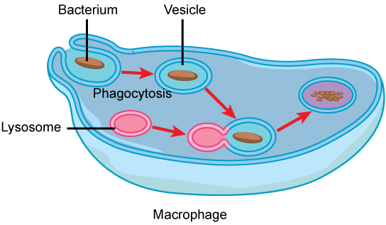
Lysosomes also use their hydrolytic enzymes to destroy disease-causing organisms that might enter the cell. A good example of this occurs in a group of white blood cells called macrophages, which are part of your body’s immune system. In a process known as phagocytosis, a section of the plasma membrane of the macrophage invaginates (folds in) and engulfs a pathogen. The invaginated section, with the pathogen inside, then pinches itself off from the plasma membrane and becomes a vesicle, sometimes called a vacuole. The vesicle fuses with a lysosome. The lysosome’s hydrolytic enzymes then destroy the pathogen (Figure 3.16).
LINK TO LEARNING
You can watch an excellent animation of the endomembrane system here. At the end of the animation, there is a short self-assessment.
Section Summary
The endomembrane system includes the nuclear envelope, lysosomes, vesicles, the ER, and Golgi apparatus, as well as the plasma membrane. These cellular components work together to modify, package, tag, and transport proteins and lipids that form the membranes.
The RER modifies proteins and synthesizes phospholipids in cell membranes. The SER synthesizes carbohydrates, lipids, and steroid hormones; engages in the detoxification of medications and poisons; and stores calcium ions. Sorting, tagging, packaging, and distributing lipids and proteins take place in the Golgi apparatus. Lysosomal enzymes are synthesized in the RER and modified in the Golgi. Lysosomes digest macromolecules, recycle worn-out organelles, and destroy pathogens.
Exercises
Glossary
endomembrane system: the group of organelles and membranes in eukaryotic cells that work together to modify, package, and transport lipids and proteins
endoplasmic reticulum (ER): a series of interconnected membranous structures within eukaryotic cells that collectively modify proteins and synthesize lipids
Golgi apparatus: a eukaryotic organelle made up of a series of stacked membranes that sorts, tags, and packages lipids and proteins for distribution
lysosome: an organelle in an animal cell that functions as the cell’s digestive component; it breaks down proteins, polysaccharides, lipids, nucleic acids, and even worn-out organelles
plasma membrane: a phospholipid bilayer with embedded (integral) or attached (peripheral) proteins that separates the internal contents of the cell from its surrounding environment
rough endoplasmic reticulum (RER): the region of the endoplasmic reticulum that is studded with ribosomes and engages in protein modification
smooth endoplasmic reticulum (SER): the region of the endoplasmic reticulum that has few or no ribosomes on its cytoplasmic surface and synthesizes carbohydrates, lipids, and steroid hormones; detoxifies chemicals like pesticides, preservatives, medications, and environmental pollutants, and stores calcium ions
vacuole: a membrane-bound sac, somewhat larger than a vesicle, that functions in cellular storage and transport
vesicle: a small, membrane-bound sac that functions in cellular storage and transport; its membrane is capable of fusing with the plasma membrane and the membranes of the endoplasmic reticulum and Golgi apparatus
Media Attributions
- Figure 3.13 modification of work by Magnus Manske
- Figure 3.14 modification of work by Louisa Howard
- Figure 3.15 modification of work by Louisa Howard; scale-bar data from Matt Russell
