3.5 The Cytoskeleton

Learning Objectives

By the end of this section, you will be able to:

  • Describe the cytoskeleton
  • Compare the roles of microfilaments, intermediate filaments, and microtubules
  • Compare and contrast cilia and flagella

Fibres of the Cytoskeleton

If you were to remove all the organelles from a cell, would the plasma membrane and the cytoplasm be the only components left? No. Within the cytoplasm, there would still be ions and organic molecules, plus a network of protein fibres that help maintain the cell’s shape, secure some organelles in specific positions, allow cytoplasm and vesicles to move within the cell, and enable cells within multicellular organisms to move. Collectively, scientists call this network of protein fibres the cytoskeleton. There are three types of fibres within the cytoskeleton: microfilaments, intermediate filaments, and microtubules (Figure 3.17). Here, we will examine each.

Figure 3.17 Microfilaments, intermediate filaments, and microtubules compose a cell’s cytoskeleton. Microfilaments thicken the cortex around the cell’s inner edge. Like rubber bands, they resist tension. There are microtubules in the cell’s interior where they maintain their shape by resisting compressive forces. There are intermediate filaments throughout the cell that hold organelles in place.

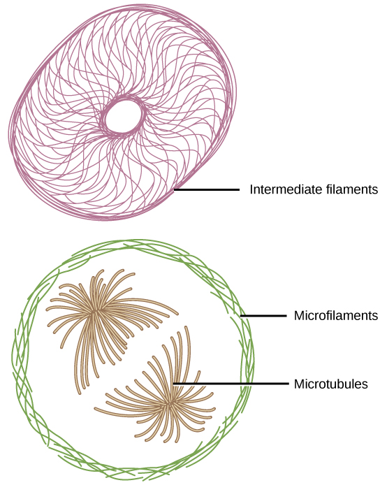
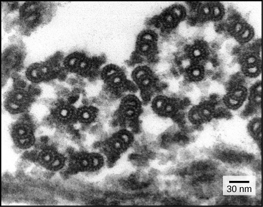
Microfilaments are the thinnest of the cytoskeletal fibres and function in moving cellular components, for example, during cell division. They also maintain the structure of microvilli, the extensive folding of the plasma membrane found in cells dedicated to absorption. These components are also common in muscle cells and are responsible for muscle cell contraction. Intermediate filaments are of intermediate diameter and have structural functions, such as maintaining the shape of the cell and anchoring organelles. Keratin, the compound that strengthens hair and nails, forms one type of intermediate filament. Microtubules are the thickest of the cytoskeletal fibres. These are hollow tubes that can dissolve and reform quickly. Microtubules guide organelle movement and are the structures that pull chromosomes to their poles during cell division. They are also the structural components of flagella and cilia. In cilia and flagella, the microtubules are organized as a circle of nine double microtubules on the outside and two microtubules in the center.

Microfilaments

Of the three types of protein fibres in the cytoskeleton, microfilaments are the narrowest. They function in cellular movement, have a diameter of about 7 nm, and are comprised of two globular protein intertwined strands, which we call actin (Figure 3.18). For this reason, we also call microfilaments actin filaments.
Figure 3.18 Two intertwined actin strands comprise microfilaments.
ATP powers actin to assemble its filamentous form, which serves as a track for the movement of a motor protein we call myosin. This enables actin to engage in cellular events requiring motion, such as cell division in eukaryotic cells and cytoplasmic streaming, which is the cell cytoplasm’s circular movement in plant cells. Actin and myosin are plentiful in muscle cells. When your actin and myosin filaments slide past each other, your muscles contract.
Microfilaments also provide some rigidity and shape to the cell. They can depolymerize (disassemble) and reform quickly, thus enabling a cell to change its shape and move. White blood cells (your body’s infection-fighting cells) make good use of this ability. They can move to an infection site and phagocytize the pathogen.

LINK TO LEARNING

To see an example of a white blood cell in action, watch a short time-lapse video of the cell capturing two bacteria. It engulfs one and then moves on to the other. Click to view content

Intermediate Filaments

Several strands of fibrous proteins that are wound together comprise intermediate filaments (Figure 3.19). Intermediate filaments get their name from the fact that their diameter, 8 to 10 nm, is between those of microfilaments and microtubules.
Figure 3.19 Intermediate filaments consist of several intertwined strands of fibrous proteins bundled together.
Intermediate filaments have no role in cell movement. Their function is purely structural. They bear tension, thus maintaining the cell’s shape, and anchor the nucleus and other organelles in place. The intermediate filaments are the most diverse group of cytoskeletal elements. Several fibrous protein types are in the intermediate filaments. You are probably most familiar with keratin, the fibrous protein that strengthens your hair, nails, and the skin’s epidermis.

Microtubules

As their name implies, microtubules are small hollow tubes. Polymerized dimers of α-tubulin and β-tubulin, two globular proteins, comprise the microtubule’s walls (Figure 3.20). With a diameter of about 25 nm, microtubules are cytoskeletons’ widest components. They help the cell resist compression, provide a track along which vesicles move through the cell, and pull replicated chromosomes to opposite ends of a dividing cell. Like microfilaments, microtubules can disassemble and reform quickly.
Figure 3.20 (Left) The microtubule’s molecular structure is a hollow cylinder. (Right) The wall of the microtubule consists of 13 polymerized dimers of α-tubulin and β-tubulin.
Microtubules are also the structural elements of flagella, cilia, and centrioles (the latter are the centrosome’s two perpendicular bodies). In animal cells, the centrosome is the microtubule-organizing center. In eukaryotic cells, flagella and cilia are quite different structurally from their counterparts in prokaryotes.

The centrosome is a region near the nucleus of animal cells and contains a pair of centrioles, two structures that lie perpendicular to each other. Each centriole is a cylinder of nine triplets of microtubules. The centrosome replicates itself before a cell divides, and the centrioles play a role in pulling the duplicated chromosomes to opposite ends of the dividing cell. However, the exact function of the centrioles in cell division is not clear, since cells that have the centrioles removed can still divide, and plant cells, which lack centrioles, are capable of cell division.

Flagella and Cilia

The flagella (singular: flagellum) are long, hair-like structures that extend from the plasma membrane and enable an entire cell to move (for example, sperm, Euglena, and some prokaryotes). When present, the cell has just one flagellum or a few flagella. However, when cilia (singular: cilium) are present, they are many in number.  Cilia extend along the plasma membrane’s entire surface. They are short, hair-like structures that move entire cells (such as paramecia) or substances along the cell’s outer surface (for example, the cilia of cells lining the Fallopian tubes that move the ovum toward the uterus, or cilia lining the cells of the respiratory tract that trap particulate matter and move it toward your nostrils).
Despite their differences in length and number, flagella and cilia share a common structural arrangement of microtubules called a “9 + 2 array.” This is an appropriate name because a single flagellum or cilium is made of a ring of nine microtubule doublets, surrounding a single microtubule doublet in the center (Figure 3.21).
Figure 3.21 This transmission electron micrograph of two flagella shows the microtubules’ 9 + 2 array: nine microtubule doublets surround a single microtubule doublet. (Credit: modification of work by Dartmouth Electron Microscope Facility, Dartmouth College; scale-bar data from Matt Russell)

Section Summary

The cytoskeleton has three different protein element types. From narrowest to widest, they are the microfilaments (actin filaments), intermediate filaments, and microtubules. Biologists often associate microfilaments with myosin. They provide rigidity and shape to the cell and facilitate cellular movements. Intermediate filaments bear tension and anchor the nucleus and other organelles in place. Microtubules help the cell resist compression, serve as tracks for motor proteins that move vesicles through the cell, and pull replicated chromosomes to opposite ends of a dividing cell. They are also the structural element of centrioles, flagella, and cilia.

Exercises

Glossary

cilium (plural: cilia): a short, hair-like structure that extends from the plasma membrane in large numbers and is used to move an entire cell or move substances along the outer surface of the cell

cytoskeleton: the network of protein fibers that collectively maintains the shape of the cell, secures some organelles in specific positions, allows cytoplasm and vesicles to move within the cell, and enables unicellular organisms to move

flagellum (plural: flagella): the long, hair-like structure that extends from the plasma membrane and is used to move the cell

Media Attribution

  • Figure 3.21 modification of work by Dartmouth Electron Microscope Facility, Dartmouth College; scale-bar data from Matt Russell

License

Icon for the Creative Commons Attribution 4.0 International License

Introduction to Biology Copyright © 2023 by Natasha Ramroop Singh, PhD is licensed under a Creative Commons Attribution 4.0 International License, except where otherwise noted.

Share This Book