3.6 Connections Between Cells
Learning Objectives
By the end of this section, you will be able to:
- Describe the extracellular matrix
- List examples of the ways that plant cells and animal cells communicate with adjacent cells
- Summarize the roles of plasmodesmata, tight junctions, gap junctions, and desmosomes
You already know that a tissue is a group of similar cells working together. As you might expect, if cells are to work together, they must communicate with each other, just as you need to communicate with others if you work on a group project. Let’s take a look at how cells communicate with each other.
Extracellular Matrix of Animal Cells
While cells in most multicellular organisms release materials into the extracellular space, animal cells will be discussed as an example. The primary components of these materials are proteins, and the most abundant protein is collagen. Collagen fibers are interwoven with proteoglycans, which are carbohydrate-containing protein molecules. Collectively, we call these materials the extracellular matrix (Figure 3.22). Not only does the extracellular matrix hold the cells together to form a tissue, but it also allows the cells within the tissue to communicate with each other. How can this happen?
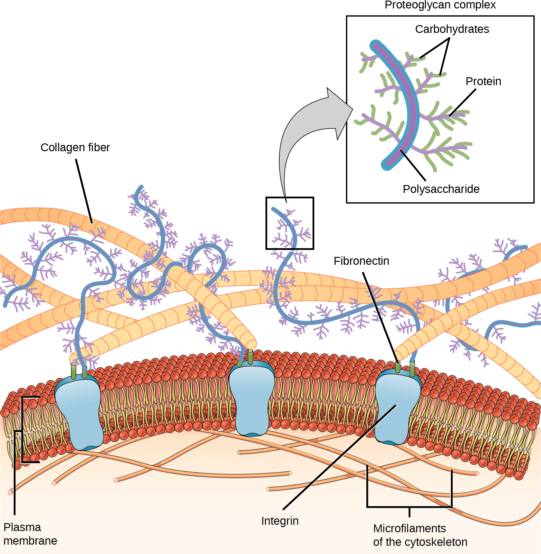
Cells have protein receptors on their plasma membranes’ extracellular surfaces. When a molecule within the matrix binds to the receptor, it changes the receptor’s molecular structure. The receptor, in turn, changes the microfilaments’ conformation positioned just inside the plasma membrane. These conformational changes induce chemical signals inside the cell that reach the nucleus and turn “on” or “off” the transcription of specific DNA sections, which affects the associated protein production, thus changing the activities within the cell.
Blood clotting provides an example of the extracellular matrix’s role in cell communication. When the cells lining a blood vessel are damaged, they display a protein receptor, which we call tissue factor. When tissue factor binds with another factor in the extracellular matrix, it causes platelets to adhere to the damaged blood vessel’s wall, stimulates the adjacent smooth muscle cells in the blood vessel to contract (thus constricting the blood vessel), and initiates a series of steps that stimulate the platelets to produce clotting factors.
Intercellular Junctions
Cells can also communicate with each other by direct contact, referred to as intercellular junctions. There are some differences in the ways that plant and animal cells do this. Plasmodesmata (singular: plasmodesma) are junctions between plant cells, whereas animal cell contacts include tight junctions, gap junctions, and desmosomes.
Plasmodesmata
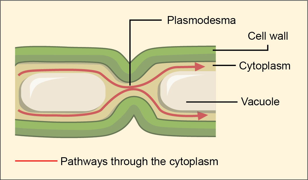
In general, long stretches of the plasma membranes of neighboring plant cells cannot touch one another because the cell wall that surrounds each cell separates them. How then, can a plant transfer water and other soil nutrients from its roots, through its stems, and to its leaves? Such transport uses the vascular tissues (xylem and phloem) primarily. There also exist structural modifications, which we call plasmodesmata (singular: plasmodesma). Plasmodesmata are numerous channels that pass between the cell walls of adjacent plant cells, connecting their cytoplasm, and enabling transport of signalling molecules and nutrients from cell to cell, and thus throughout the plant (Figure 3.23).
Tight Junctions
A tight junction is a watertight seal between two adjacent animal cells (Figure 3.24). Proteins, predominantly claudins and occludins, tightly hold the cells against each other. This tight adherence prevents materials from leaking between the cells; tight junctions are typically found in epithelial tissues that line internal organs and cavities, and comprise most of the skin. For example, the tight junctions of the epithelial cells lining your urinary bladder prevent urine from leaking out into the extracellular space.

Gap Junctions
Gap junctions in animal cells are like plasmodesmata in plant cells in that they are channels between adjacent cells that allow for transporting ions, nutrients, and other substances that enable cells to communicate (Figure 3.25). Structurally, however, gap junctions and plasmodesmata differ.

Gap junctions develop when a set of six proteins, connexins, in the plasma membrane arrange themselves in an elongated doughnut-like configuration to form a connexon. When the connexon’s pores (“doughnut holes”) in adjacent animal cells align, a channel between the two cells forms. Gap junctions are particularly important in cardiac muscle. The electrical signal for the muscle to contract passes efficiently through gap junctions, allowing the heart muscle cells to contract in tandem.
CAREER CONNECTION
Cardiologist
Heart disease is the leading cause of death in the United States. This is primarily due to our sedentary lifestyle and our high trans-fat diets.
Heart failure is just one of many disabling heart conditions. Heart failure does not mean that the heart has stopped working. Rather, it means that the heart can’t pump with sufficient force to transport oxygenated blood to all the vital organs. Left untreated, heart failure can lead to kidney failure and other organ failure.
Cardiac muscle tissue comprises the heart’s wall. Heart failure occurs when cardiac muscle cells’ endoplasmic reticula do not function properly. As a result, an insufficient number of calcium ions are available to trigger a sufficient contractile force.
Cardiologists (cardi- = “heart”; -ologist = “one who studies”) are doctors who specialize in treating heart diseases, including heart failure. Cardiologists can diagnose heart failure via a physical examination, results from an electrocardiogram (ECG, a test that measures the heart’s electrical activity), a chest X-ray to see whether the heart is enlarged, and other tests. If the cardiologist diagnoses heart failure, he or she will typically prescribe appropriate medications and recommend a reduced table salt intake and a supervised exercise program.
Desmosomes
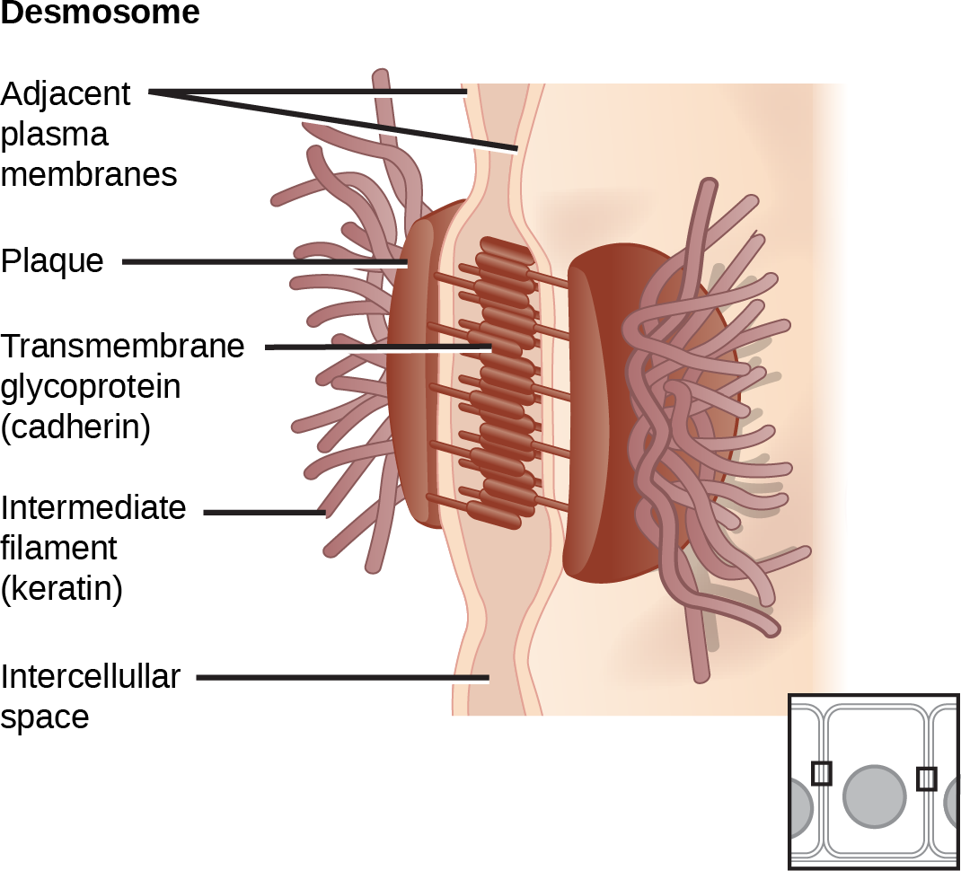
Only animal cells have desmosomes, which act like spot welds between adjacent epithelial cells (Figure 3.26). Cadherins, short proteins in the plasma membrane, connect to intermediate filaments to create desmosomes. The cadherins connect two adjacent cells and maintain the cells in a sheet-like formation in organs and tissues that stretch, like the skin, heart, and muscles.
Section Summary
Animal cells communicate via their extracellular matrices and are connected to each other via tight junctions, desmosomes, and gap junctions. Plant cells are connected and communicate with each other via plasmodesmata. When protein receptors on the plasma membrane’s surface of an animal cell bind to a substance in the extracellular matrix, a chain of reactions begins that changes activities taking place within the cell. Plasmodesmata are channels between adjacent plant cells, while gap junctions are channels between adjacent animal cells. However, their structures are quite different. A tight junction is a watertight seal between two adjacent cells, while a desmosome acts like a spot weld.
Exercises
Glossary
desmosome: a linkage between adjacent epithelial cells that forms when cadherins in the plasma membrane attach to intermediate filaments
extracellular matrix: the material, primarily collagen, glycoproteins, and proteoglycans, secreted from animal cells that holds cells together as a tissue, allows cells to communicate with each other, and provides mechanical protection and anchoring for cells in the tissue
gap junction: a channel between two adjacent animal cells that allows ions, nutrients, and other low-molecular weight substances to pass between the cells, enabling the cells to communicate
plasmodesma (plural: plasmodesmata): a channel that passes between the cell walls of adjacent plant cells, connects their cytoplasm, and allows materials to be transported from cell to cell
tight junction: a firm seal between two adjacent animal cells created by protein adherence
Media Attributions
- Figure 3.24 modification of work by Mariana Ruiz Villareal
- Figure 3.25 modification of work by Mariana Ruiz Villareal
- Figure 3.26 modification of work by Mariana Ruiz Villareal
