3.7 Mitochondria and Chloroplasts
Learning Objectives
By the end of this section, you will be able to:
- Describe the structures and functions of mitochondria and chloroplasts
- Describe the theory of endosymbiosis
Mitochondria
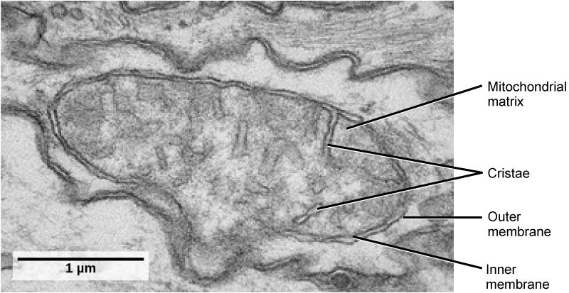
Mitochondria (singular: mitochondrion) are often called the “powerhouses” or “energy factories” of a cell because they are responsible for making ATP, the cell’s main energy-carrying molecule. The formation of ATP from the breakdown of glucose is known as cellular respiration. Mitochondria are oval-shaped, double-membrane organelles (Figure 3.27) that have their own ribosomes and DNA. Each membrane is a phospholipid bilayer embedded with proteins. The inner membrane has folds called cristae, which increase the surface area of the inner membrane. The area surrounded by the folds is called the mitochondrial matrix. The cristae and the matrix have different roles in cellular respiration.
Chloroplasts
Like mitochondria, chloroplasts also have their own DNA and ribosomes. Chloroplasts function in photosynthesis and can be found in eukaryotic cells such as plants and algae. In photosynthesis, carbon dioxide, water, and light energy are used to make glucose and oxygen. This is the major difference between plants and animals: plants (autotrophs) are able to make their own food, like glucose, whereas animals (heterotrophs) must rely on other organisms for their organic compounds or food source.
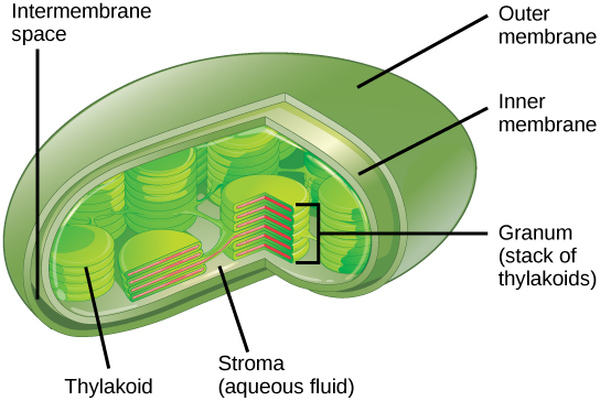
Like mitochondria, chloroplasts have outer and inner membranes, but within the space enclosed by a chloroplast’s inner membrane is a set of interconnected and stacked, fluid-filled membrane sacs called thylakoids (Figure 3.28). Each stack of thylakoids is called a granum (plural: grana). The fluid enclosed by the inner membrane and surrounding the grana is called the stroma.
The chloroplasts contain a green pigment called chlorophyll, which captures the energy of sunlight for photosynthesis. Like plant cells, photosynthetic protists also have chloroplasts. Some bacteria also perform photosynthesis, but they do not have chloroplasts. Their photosynthetic pigments are located in the thylakoid membrane within the cell itself.
Watch a video about how the evolution of organ systems and organisms may be influenced by mitochondria and chloroplasts living inside eukaryotic cells.
Endosymbiosis
We have mentioned that both mitochondria and chloroplasts contain DNA and ribosomes. Have you wondered why? Strong evidence points to endosymbiosis as the explanation.
Symbiosis is a relationship in which organisms from two separate species live in close association and typically exhibit specific adaptations to each other. Endosymbiosis (endo- = “within”) is a relationship in which one organism lives inside the other. Endosymbiotic relationships abound in nature. Microbes that produce vitamin K live inside the human gut. This relationship is beneficial for us because we are unable to synthesize vitamin K. It is also beneficial for the microbes because they are protected from other organisms and are provided a stable habitat and abundant food by living within the large intestine.
Scientists have long noticed that bacteria, mitochondria, and chloroplasts are similar in size. We also know that mitochondria and chloroplasts have DNA and ribosomes, just as bacteria do and they resemble the types found in bacteria. Scientists believe that host cells and bacteria formed a mutually beneficial endosymbiotic relationship when the host cells ingested aerobic bacteria and cyanobacteria but did not destroy them. Through evolution, these ingested bacteria became more specialized in their functions, with the aerobic bacteria becoming mitochondria and the photosynthetic bacteria becoming chloroplasts.
Section Summary
Eukaryotic cells produce the majority of their ATP in mitochondria. The inner mitochondrial membrane is folded into cristae surrounding the matrix. Plant cells have mitochondria to produce ATP but also chloroplasts to generate glucose by photosynthesis.
The endosymbiotic theory suggests that mitochondria and chloroplasts were originally prokaryotic cells that had been endocytosed into what are now eukaryotic cells. Mitochondria and chloroplasts contain their own DNA and their ribosomes resemble those of bacteria.
Exercises
Glossary
chloroplast: a plant cell organelle that carries out photosynthesis
mitochondria (singular: mitochondrion): the cellular organelles responsible for carrying out cellular respiration, resulting in the production of ATP, the cell’s main energy-carrying molecule
Media Attribution
- Figure 3.27 modification of work by Matthew Britton; scale-bar data from Matt Russell)
